Veterinary Laboratory & Diagnostics in Matthews
State-of-the-Art Diagnostic Imaging: Pet MRI, CT Scans, and Ultrasounds
At Carolina Veterinary Specialists in Matthews, our veterinarians and board-certified specialists use in-house diagnostic tests and tools such as pet MRI, CT scans, and ultrasounds to provide the most accurate diagnosis of medical conditions and tailor treatment plans to the needs of your pet.
Diagnosis & Treatment Planning
Diagnostic imaging is the use of electromagnetic radiation and other technologies to produce highly detailed images of the internal structures of your pet's body.
At Carolina Veterinary Specialists in Matthews, we use the most advanced tools available to help provide an accurate diagnosis of your pets’ medical issues.
Our diagnostic imaging capabilities allow us to provide more immediate treatment options, and to share information with your primary care veterinarian in the most time-efficient manner. We also accept referrals for outpatient ultrasounds from your primary care veterinarian.

In-House Lab & Pharmacy
Our in-house laboratory allows us to perform tests and get results quickly, so that we can diagnose your pet's symptoms and start treatment as soon as possible. Our pharmacy is stocked with a range of medications and prescription diets, giving us quick access to any medications your pets may need while in our care.
Our Diagnostic Services: Pet MRIs, CT Scans and Ultrasounds
At Carolina Veterinary Specialists in Matthews, we are pleased to offer advanced diagnostics which include a Pet MRI machine, a CT scanner and ultrasound.
These tools allow our specialists and veterinarians to provide an accurate diagnosis of your pets’ medical issues.
- Radiography (Digital X-Rays)
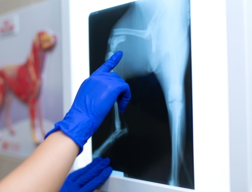
A radiograph, or digital x-ray, is a type of photograph that can look inside the body and reveal information that may not be discernible from the outside.
Radiography is painless, safe, and completely non-invasive, and uses only very low doses of radiation. Because the level of radiation exposure needed to perform radiography is very low, even pregnant females and very young pets can undergo radiography.
Radiographs can be used to evaluate bones and organs and diagnose conditions including bladder stones, broken bones, chronic arthritis, spinal cord diseases and some tumors. - Ultrasound
Ultrasound imaging involves exposing part of the body to high-frequency sound waves to produce pictures of the inside of the body.
Because ultrasound images are captured in real-time, they can show the structure and movement of the body's internal organs, as well as blood flowing through blood vessels.
- Echocardiography
Echocardiography is a non-invasive test that allows your veterinary cardiologist to directly evaluate the anatomy and function of the heart.
An echocardiogram is an ultrasound image of the heart and associated large blood vessels.
- CT Scan (Computed Tomography)
A CT scanner is an advanced, non-invasive diagnostic machine that allows our specialists to view the internal anatomy of a pet by producing highly detailed cross-sectional images.
CT scans are commonly used for planning surgery, diagnosing diseases and the early detection and treatment of cancer.
- MRI (Magnetic Resonance Imaging)
Magnetic resonance imaging (MRI) uses a powerful magnetic field, radio waves and a computer to produce detailed pictures of organs, soft tissues, and other internal body structures.
The images can then be examined on a computer monitor or printed.
- Endoscopy
An endoscope is made up of a very tiny camera and light affixed to the end of a flexible tube.
An endoscopy is the insertion of a long, thin tube directly into the body to observe an internal organ or tissue in detail.
Endoscopes are minimally invasive and can be inserted into the openings of the body such as the mouth or anus.


